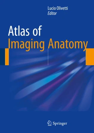
Atlas of Imaging Anatomy PDF – Comprehensive Reference for Radiology and Anatomy
The Atlas of Imaging Anatomy PDF is an essential reference that combines high-quality imaging with detailed anatomical illustrations, offering clinicians, radiologists, and medical students a complete guide to cross-sectional anatomy. With updated imaging modalities and clinically relevant perspectives, this atlas provides a strong foundation for understanding normal structures and their variations. It is a trusted resource for both diagnostic imaging and academic learning.
Why This Book Matters
Accurate interpretation of imaging studies requires a solid understanding of anatomy in different modalities such as CT, MRI, and ultrasound. Atlas of Imaging Anatomy delivers clear, labeled images that bridge the gap between textbook anatomy and clinical imaging. It enables faster recognition of anatomical landmarks, supports accurate diagnoses, and enhances communication among healthcare professionals.
For related updates and resources in radiology, visit the Radiological Society of North America (RSNA) and the European Society of Radiology (ESR).
Key Features of the Ebook
This atlas includes:
-
High-resolution imaging correlated with detailed anatomical drawings
-
Coverage of CT, MRI, X-ray, and ultrasound anatomy
-
Systematic organization by body region for easy reference
-
Clinically relevant notes and variations
-
Essential cross-sectional imaging for head, chest, abdomen, pelvis, and extremities
-
Educational value for both training and clinical practice
For additional authoritative sources, explore the Journal of Radiology and the American Journal of Roentgenology (AJR).
Who Can Benefit
This ebook is designed for:
-
Radiologists and radiology residents
-
Medical students and anatomy learners
-
Surgeons and clinicians interpreting imaging
-
Emergency physicians using cross-sectional imaging
-
Educators and researchers in medical imaging
For complementary study, consider resources like Pocket Atlas of Sectional Anatomy and Learning Radiology: Recognizing the Basics.
Learning and Application Strategies
The Atlas of Imaging Anatomy emphasizes correlation between imaging and gross anatomy, making it a practical study and clinical reference. By combining annotated images with concise explanations, it supports students preparing for exams and clinicians seeking quick reference during interpretation. Its structured format ensures easy access to essential anatomical details across different imaging techniques.
For further resources, consult the American College of Radiology (ACR) and the Society of Abdominal Radiology (SAR).
Detailed Content Overview
Chapters are organized to cover:
-
Head and neck imaging anatomy
-
Thoracic and cardiovascular system anatomy
-
Abdominal and pelvic structures
-
Musculoskeletal imaging of upper and lower extremities
-
Neuroimaging with CT and MRI correlation
-
Ultrasound and cross-sectional perspectives
-
Annotated charts, labels, and quick-reference diagrams
Conclusion
The Atlas of Imaging Anatomy PDF is a must-have reference for understanding cross-sectional anatomy across imaging modalities. By integrating detailed illustrations with radiological images, it enhances diagnostic accuracy, supports academic learning, and improves clinical communication.
👉 Download Atlas of Imaging Anatomy PDF today to advance your radiology and anatomy knowledge. For further resources, visit FreeMedBooks and explore purchase options on Amazon.

