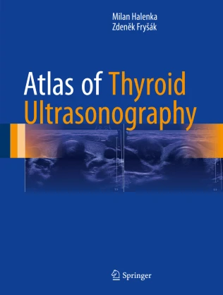
Atlas of Thyroid Ultrasonography PDF – Comprehensive Imaging Reference for Thyroid Disorders
The Atlas of Thyroid Ultrasonography PDF is a detailed, evidence-based guide designed to support clinicians in the diagnosis and management of thyroid diseases through ultrasound imaging. Written by experts in endocrinology and radiology, this atlas provides high-quality images, structured explanations, and practical guidelines that are essential for accurate evaluation of thyroid nodules, tumors, and other pathologies. Compact and well-organized, it is a valuable resource for endocrinologists, radiologists, surgeons, and medical students.
Why This Book Matters
Thyroid ultrasonography plays a central role in the detection and follow-up of thyroid nodules, goiter, and malignancies. Accurate interpretation of ultrasound features is critical for guiding fine-needle aspiration (FNA), risk stratification, and treatment planning. The Atlas of Thyroid Ultrasonography provides clinicians with standardized imaging criteria, ensuring better diagnostic accuracy and improved patient care.
For guidelines and updates in thyroid imaging, visit the American Thyroid Association (ATA) and the European Thyroid Association (ETA).
Key Features of the Ebook
This comprehensive atlas includes:
-
High-resolution ultrasound images of thyroid nodules and pathologies
-
Detailed explanations of sonographic features and classification systems
-
Guidance for fine-needle aspiration biopsy (FNAB) techniques
-
Risk stratification using TI-RADS and ATA guidelines
-
Case-based examples for clinical practice
-
Updates on thyroid cancer imaging and management
-
Practical charts and diagnostic algorithms for rapid reference
For further resources, consult the Journal of Clinical Endocrinology & Metabolism (JCEM) and Radiology Journal.
Who Can Benefit
This ebook is designed for:
-
Endocrinologists and radiologists
-
Surgeons managing thyroid and parathyroid diseases
-
Pathologists involved in thyroid cytology
-
Medical students and residents in imaging specialties
-
Clinicians seeking reliable imaging guidance for thyroid disorders
For complementary resources, explore Thyroid Cancer: A Comprehensive Guide to Clinical Management and Diagnostic Ultrasound: Head and Neck.
Learning and Application Strategies
The atlas emphasizes practical interpretation of ultrasound findings with annotated images and structured reporting methods. By combining classification systems, biopsy guidelines, and imaging criteria, it equips clinicians to provide precise and evidence-based care in both routine and complex thyroid cases. Its visual approach ensures faster learning and enhanced diagnostic confidence.
For additional resources, visit the World Health Organization (WHO) endocrine tumor classification and the Endocrine Society.
Detailed Content Overview
Chapters are organized to cover:
-
Normal thyroid anatomy and ultrasound landmarks
-
Classification of benign and malignant thyroid nodules
-
TI-RADS, ATA, and other risk stratification systems
-
Fine-needle aspiration techniques and indications
-
Imaging of thyroid cancer and postoperative surveillance
-
Parathyroid imaging and related disorders
-
Essential charts, diagnostic tables, and case illustrations
Conclusion
The Atlas of Thyroid Ultrasonography PDF is a definitive imaging reference that combines high-quality sonographic visuals with practical clinical guidance. By offering standardized diagnostic criteria and risk stratification tools, it supports clinicians in delivering accurate and effective thyroid care.
👉 Download Atlas of Thyroid Ultrasonography PDF today to improve your diagnostic skills and patient outcomes. For further access, explore FreeMedBooks and purchase a copy on Amazon.

