
Cranial Neuroimaging and Clinical Neuroanatomy 4E PDF – Comprehensive Atlas for MR Imaging and CT
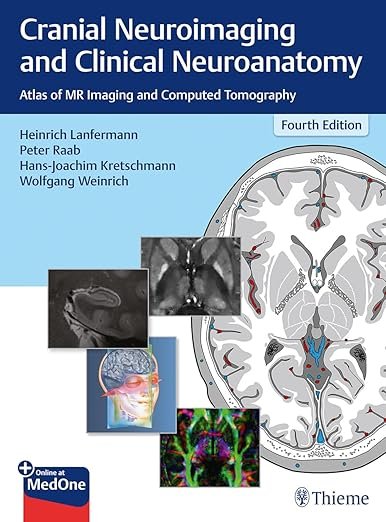
The Cranial Neuroimaging and Clinical Neuroanatomy: Atlas of MR Imaging and Computed Tomography 4e PDF is a highly acclaimed reference that integrates detailed neuroanatomy with advanced imaging techniques. Written by leading experts in neuroradiology and clinical neuroscience, this atlas provides clinicians with an authoritative guide to interpreting both MRI and CT images of the brain and cranial structures. With its updated content and high-quality illustrations, it is an indispensable tool for radiologists, neurologists, neurosurgeons, and medical students.
Why This Book Matters
Accurate interpretation of neuroimaging is essential for diagnosing neurological disorders, planning surgical interventions, and guiding patient care. This atlas combines clear anatomical illustrations with practical imaging examples, ensuring precise correlation between structure and pathology. The 4th edition reflects the latest developments in MR and CT imaging, making it a vital reference for modern clinical practice.
For current guidelines and updates in neuroradiology, visit the Radiological Society of North America (RSNA) and the American Society of Neuroradiology (ASNR).
Key Features of the Ebook
This atlas includes:
-
High-resolution MR and CT images with detailed annotations
-
Correlation between cross-sectional anatomy and imaging findings
-
Coverage of cranial nerves, brainstem, and vascular structures
-
Pathological imaging examples for clinical relevance
-
Updated techniques and imaging protocols in neuroimaging
-
Concise tables and diagrams for quick reference
For complementary research, consult the Journal of Neuroradiology and the European Society of Neuroradiology (ESNR).
Who Can Benefit
This ebook is designed for:
-
Radiologists and neuroradiologists
-
Neurologists and neurosurgeons
-
Medical students and residents in radiology or neurology
-
Clinicians seeking a reliable imaging atlas for diagnosis
-
Researchers in neuroimaging and neuroanatomy
For additional resources, explore Duus’ Topical Diagnosis in Neurology and MRI of the Brain: A Teaching Atlas.
Learning and Application Strategies
The book emphasizes direct clinical application, allowing readers to correlate anatomical knowledge with imaging findings quickly. By integrating MR and CT images with labeled structures, the atlas enhances diagnostic accuracy and supports efficient decision-making in neurology and neurosurgery. Its structured format makes it ideal for both study and clinical practice.
For further educational resources, visit the American College of Radiology (ACR) and the National Center for Biotechnology Information (NCBI).
Detailed Content Overview
Chapters are organized to cover:
-
Fundamentals of neuroanatomy and imaging
-
Cross-sectional anatomy with MR and CT correlation
-
Cranial nerves and brainstem anatomy
-
Cerebral vasculature and pathology
-
Imaging of common neurological disorders
-
Pediatric neuroimaging essentials
-
Clinical case studies with annotated images
Conclusion
The Cranial Neuroimaging and Clinical Neuroanatomy: Atlas of MR Imaging and Computed Tomography 4e PDF is a trusted and comprehensive resource for mastering neuroimaging interpretation. With its detailed anatomical guidance and high-quality imaging, it empowers clinicians and students to improve diagnostic precision and clinical outcomes.
👉 Download Cranial Neuroimaging and Clinical Neuroanatomy 4E PDF today to enhance your neuroimaging expertise. For more references, explore FreeMedBooks and purchase the book on Amazon.