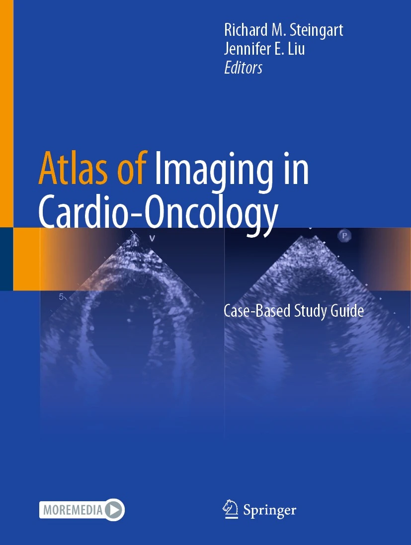
Atlas of Imaging in Cardio-Oncology PDF
The Atlas of Imaging in Cardio-Oncology PDF is a comprehensive guide for cardiologists, oncologists, radiologists, and healthcare professionals. This atlas provides detailed coverage of cardiovascular imaging in cancer patients, highlighting the detection, assessment, and management of cardiotoxicity caused by cancer therapies. With high-quality images, illustrations, and case-based examples, it helps clinicians optimize patient care and improve outcomes.
Why Cardio-Oncology Imaging Matters
Cardiovascular complications are common in patients undergoing cancer therapy. Early and accurate imaging is critical for detecting cardiotoxicity, assessing cardiac function, and guiding interventions. Understanding imaging modalities and interpretation ensures timely diagnosis, prevents long-term complications, and improves patient prognosis. For more information, visit the American Heart Association and the Society for Cardiovascular Magnetic Resonance.
Key Features of the Ebook
This book provides:
Comprehensive coverage of cardiovascular imaging techniques in oncology patients
High-quality images and detailed illustrations for better understanding
Case-based examples demonstrating real-world diagnostic and management scenarios
Guidance on interpretation, clinical decision-making, and monitoring strategies
Evidence-based recommendations for follow-up and risk assessment
For updated clinical protocols, see the Radiological Society of North America.
Who Can Benefit
This atlas is ideal for cardiologists, oncologists, radiologists, fellows, residents, and other healthcare professionals involved in cardio-oncology care. It enhances diagnostic accuracy, supports clinical decision-making, and improves patient safety.
Integrated Learning Approach
The ebook combines theoretical principles, imaging interpretation, and practical case studies. Clinicians can apply these insights directly to patient care, ensuring accurate diagnosis, effective monitoring, and timely interventions. This integrated approach supports professional development in cardio-oncology and cardiovascular imaging.
Detailed Content Overview
The content includes:
Cardiovascular complications of cancer therapy
Imaging modalities including echocardiography, cardiac CT, MRI, and nuclear techniques
Assessment of cardiac function, structure, and tissue characterization
Monitoring cardiotoxicity and risk stratification
Case-based examples illustrating imaging interpretation and clinical decision-making
Guidance for intervention planning and patient follow-up
For additional resources, visit the Society for Cardiovascular Magnetic Resonance and the American Heart Association.
Conclusion
The Atlas of Imaging in Cardio-Oncology PDF is an essential resource for clinicians managing cardiovascular care in cancer patients. With structured guidance, high-quality imaging, and case-based learning, it equips healthcare professionals to make accurate diagnoses and deliver optimal patient care. Access this ebook today to enhance your expertise in cardio-oncology imaging.
For official educational resources, visit the American College of Cardiology.
