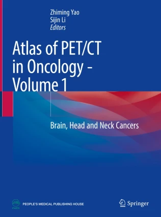
Atlas of PET/CT in Oncology – Volume 1: Brain, Head and Neck Cancers PDF – Essential Imaging Reference
The Atlas of PET/CT in Oncology – Volume 1: Brain, Head and Neck Cancers PDF is a comprehensive and authoritative reference designed to guide clinicians in the accurate interpretation of PET/CT imaging for oncology cases. Focused on brain, head, and neck cancers, this volume integrates detailed case studies, high-quality PET/CT images, and clinical insights, making it an indispensable tool for radiologists, oncologists, and nuclear medicine specialists. With a clear and structured format, it serves both as a teaching guide and as a quick reference for clinical practice.
Why This Book Matters
Oncologic imaging requires precise differentiation between tumor recurrence, treatment effects, and benign findings. PET/CT has become one of the most reliable imaging modalities in oncology, providing functional and anatomical information in a single scan. This atlas delivers practical guidance on image interpretation, clinical correlations, and treatment planning. By offering case-based examples, it enhances diagnostic accuracy and improves multidisciplinary cancer care.
For authoritative oncology imaging guidelines, visit the European Association of Nuclear Medicine (EANM) and the American Cancer Society (ACS).
Key Features of the Ebook
This atlas provides:
-
Extensive PET/CT imaging cases of brain, head, and neck cancers
-
High-resolution images with annotations for accurate interpretation
-
Diagnostic pearls to distinguish malignant vs. benign findings
-
Integration of clinical data with imaging results
-
Guidelines for staging, restaging, and therapy assessment
-
Educational insights for trainees and experienced clinicians alike
-
Updated oncologic imaging standards and best practices
For additional references, explore the Journal of Nuclear Medicine (JNM) and the National Cancer Institute (NCI).
Who Can Benefit
This ebook is designed for:
-
Radiologists and nuclear medicine specialists
-
Oncologists and radiation oncologists
-
Medical physicists and imaging technologists
-
Residents and fellows in oncology and radiology
-
Clinicians seeking advanced reference for oncologic PET/CT
For complementary resources, consult Clinical PET-CT in Radiology and PET/CT in Head and Neck Cancer.
Learning and Application Strategies
The book emphasizes real-world application of PET/CT imaging through carefully selected cases. Each chapter includes clinical context, scan interpretation, and key diagnostic considerations. By combining anatomical and metabolic imaging findings, clinicians can optimize cancer detection, treatment planning, and follow-up strategies. The concise atlas format allows for rapid access during clinical practice or academic study.
For more educational oncology imaging materials, visit the Radiological Society of North America (RSNA) and the American Society of Clinical Oncology (ASCO).
Detailed Content Overview
Chapters are organized to cover:
-
PET/CT principles and oncology imaging fundamentals
-
Brain tumors and differential diagnosis with PET/CT
-
Imaging of head and neck cancers with clinical correlations
-
Post-treatment evaluation and recurrence detection
-
Common pitfalls and artifacts in PET/CT interpretation
-
Case-based learning with annotated images
-
Practical clinical guidelines and reference tables
Conclusion
The Atlas of PET/CT in Oncology – Volume 1: Brain, Head and Neck Cancers PDF is an essential reference for accurate and clinically relevant oncologic imaging. With its combination of detailed images, case discussions, and practical guidance, it equips clinicians with the tools necessary to enhance cancer diagnosis and patient management.
👉 Download Atlas of PET/CT in Oncology – Volume 1: Brain, Head and Neck Cancers PDF today to advance your oncology imaging practice. For more details, explore FreeMedBooks and purchase the official edition on Amazon.

