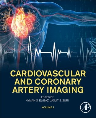
Cardiovascular and Coronary Artery Imaging Volume 2 PDF
FREE
Cardiovascular and Coronary Artery Imaging Volume 2 PDF
Introduction
Cardiovascular and Coronary Artery Imaging Volume 2 builds upon the foundation of Volume 1, offering deeper insights into advanced imaging applications in complex cardiovascular conditions. This volume focuses on specialized techniques, case-based applications, and cutting-edge technologies that shape modern cardiac diagnostics and therapy planning. According to the American Heart Association (AHA), advanced imaging improves diagnostic precision, guides interventional procedures, and enhances patient outcomes in coronary artery disease and heart failure.
Why Advanced Imaging Matters in Modern Cardiology
While basic imaging modalities provide essential diagnostic information, advanced cardiovascular imaging allows clinicians to detect subclinical disease, evaluate myocardial function, and guide high-risk interventions. The National Institutes of Health (NIH) emphasizes that multimodality imaging is essential in tailoring therapy to the individual patient, particularly in complex or refractory cases.
Specialized Imaging Techniques Covered in Volume 2
Cardiac CT and MRI in Complex Cases
Used for evaluating congenital heart disease, cardiomyopathies, and myocardial viability before surgical or interventional procedures.
Coronary Plaque Characterization
Advanced CT and MRI techniques identify vulnerable plaques that carry a high risk of rupture and myocardial infarction.
Hybrid Imaging (PET-CT, PET-MRI)
Combines functional and anatomical data to assess perfusion, metabolism, and structure in a single exam.
Stress Imaging
Dobutamine stress echocardiography, stress MRI, and nuclear stress tests are critical for detecting ischemia and functional reserve.
3D and 4D Imaging
Emerging modalities allow real-time visualization of cardiac motion, valve function, and blood flow dynamics.
Risk Stratification Through Imaging
Advanced imaging enhances patient risk assessment by identifying:
-
High-risk coronary plaques
-
Early signs of ischemia not visible in standard testing
-
Myocardial fibrosis and scarring
-
Microvascular dysfunction
-
Complex congenital abnormalities
-
Predictors of sudden cardiac death
Diagnosis and Monitoring
Volume 2 emphasizes integrating imaging into both acute and chronic cardiovascular care. Imaging is pivotal in evaluating chest pain syndromes, monitoring heart failure progression, guiding device implantation, and following up on revascularization procedures. Combining imaging with biomarkers and genetic testing improves personalized medicine approaches.
Management and Treatment
Prevention and Early Detection
Non-invasive imaging reveals preclinical atherosclerosis, enabling early lifestyle and pharmacologic interventions.
Interventional Cardiology
Intravascular imaging guides stent placement, lesion assessment, and treatment of complex coronary anatomies.
Surgical Planning
CT and MRI provide detailed anatomical maps for valve surgery, bypass grafting, and congenital defect repairs.
Multidisciplinary Collaboration
Cardiologists, radiologists, and surgeons rely on shared imaging data to make precise therapeutic decisions. The European Association of Cardiovascular Imaging (EACVI) recommends integrating multimodality imaging into routine practice to improve long-term outcomes.
Conclusion
Cardiovascular and Coronary Artery Imaging Volume 2 expands the clinical applications of imaging, offering practical knowledge for managing complex and high-risk cardiovascular patients. With its emphasis on advanced technologies and case-based learning, this volume is an indispensable guide for cardiologists, radiologists, and healthcare professionals engaged in cardiovascular care.
👉 Download Cardiovascular and Coronary Artery Imaging Volume 2 PDF to explore advanced imaging protocols, clinical case studies, and expert guidelines for precision cardiology.
For further insights, visit American Heart Association and European Association of Cardiovascular Imaging

