Overview
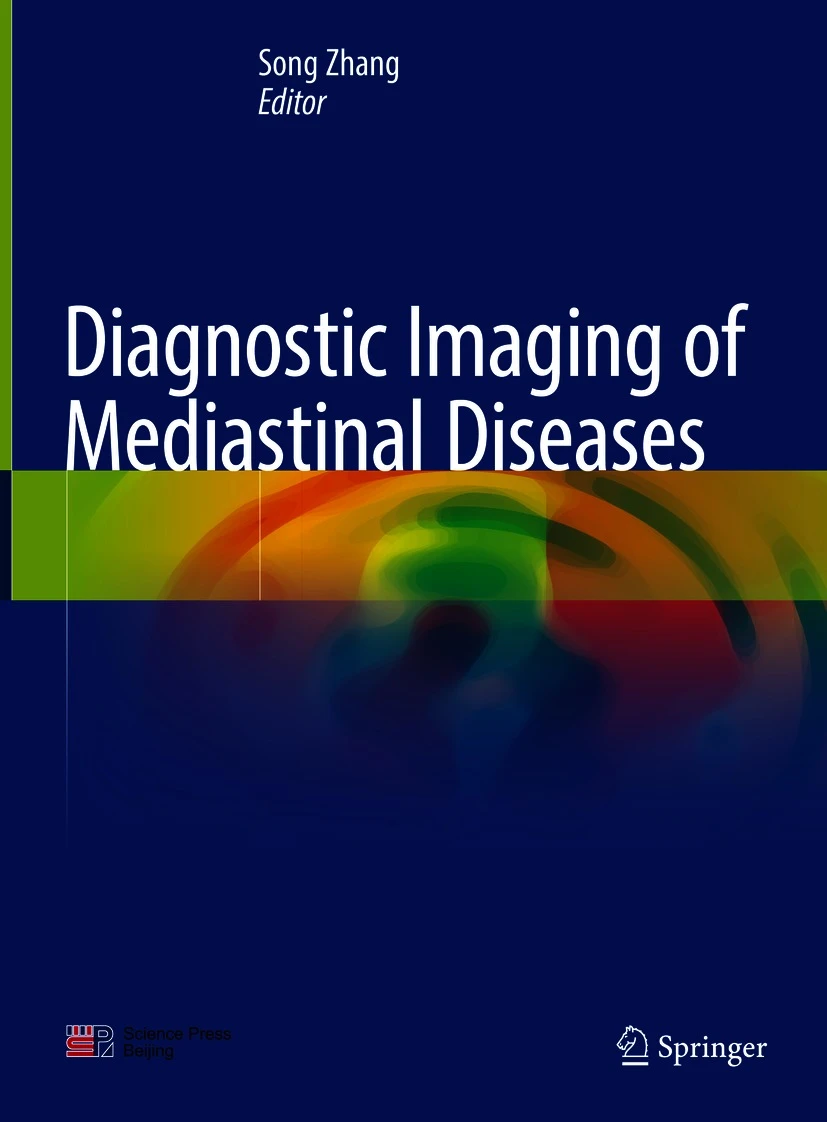
Diagnostic Imaging of Mediastinal Diseases PDF is a specialized medical reference offering comprehensive insights into imaging techniques, interpretation, and clinical applications related to mediastinal disorders. Authored by leading experts in diagnostic radiology, this book combines high-quality imaging with clear explanations, helping clinicians, radiologists, and residents improve diagnostic accuracy. Moreover, with its integration of advanced imaging modalities such as CT, MRI, and PET, it serves as an essential guide for understanding mediastinal anatomy and pathology.
Why This Book Matters
The mediastinum is a complex anatomical region containing vital structures such as the heart, great vessels, thymus, trachea, and lymphatic tissue. Accurate diagnosis of diseases in this region is challenging due to overlapping symptoms and imaging findings. Therefore, this book equips clinicians with detailed imaging strategies, differential diagnoses, and clinical correlations to approach these challenges systematically. Furthermore, it provides evidence-based insights into benign and malignant conditions, offering practical recommendations that enhance patient management. By combining detailed imaging analysis with clinical perspectives, it bridges the gap between radiology and patient-centered care.
For additional professional resources, you can visit the Radiological Society of North America (RSNA) or explore thoracic imaging guidelines at the American College of Radiology (ACR).
Key Features
This reference includes:
Comprehensive Coverage
Detailed explanations of mediastinal anatomy, pathophysiology, and disease classification provide the foundation for accurate diagnosis.
Advanced Imaging Protocols
Step-by-step guidance for CT, MRI, and PET ensures precise evaluation and consistent results.
High-Resolution Illustrations
The book features radiologic images, diagrams, and case-based examples that clarify complex findings.
Evidence-Based Discussions
Comparisons of benign and malignant conditions allow readers to understand controversies and apply best practices.
Practical Clinical Value
Tips for differential diagnosis, avoidance of pitfalls, and complication management make it highly applicable to real-world practice.
In addition, the combination of illustrative cases and practical recommendations ensures relevance for radiologists, pulmonologists, oncologists, and surgeons.
Who Can Benefit
This book is particularly valuable for:
-
Radiologists specializing in thoracic and mediastinal imaging
-
Residents and fellows in diagnostic radiology and thoracic surgery programs
-
Pulmonologists and oncologists managing complex mediastinal diseases
-
Surgeons requiring detailed imaging guidance for operative planning
-
Medical and academic libraries supporting radiology education
Consequently, it serves as both a training reference and a clinical decision-making tool.
Content Overview
The book covers:
-
Anatomy and imaging characteristics of the mediastinum
-
Protocols for CT, MRI, and PET imaging in mediastinal evaluation
-
Differential diagnosis of mediastinal masses and lymphadenopathy
-
Imaging features of thymic, vascular, and congenital anomalies
-
Diagnostic strategies for malignant mediastinal tumors
-
Case-based discussions integrating imaging findings with clinical management
-
Post-treatment imaging evaluation and long-term follow-up
Moreover, by emphasizing practical application, the book enables clinicians to translate imaging findings into effective patient care decisions.
Conclusion
Diagnostic Imaging of Mediastinal Diseases PDF is an authoritative and practical resource for mastering mediastinal imaging. By combining state-of-the-art imaging techniques with clinical context, it empowers healthcare professionals to achieve precise diagnoses and optimize treatment strategies. Therefore, it stands out as an indispensable tool for both practicing radiologists and trainees.
🔗 Download from Freemedbooks.com
🔗 Purchase on Amazon.com

