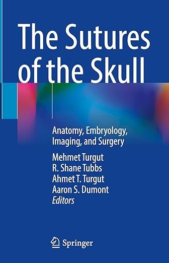
The Sutures of the Skull: Anatomy, Embryology, Imaging, and Surgery PDF
FREE
The Sutures of the Skull: Anatomy, Embryology, Imaging, and Surgery PDF is a unique, specialized reference that provides a comprehensive overview of cranial sutures, from developmental biology to advanced imaging and surgical applications. Written by leading experts, this book integrates anatomical details, embryological development, radiological evaluation, and clinical surgical approaches, making it an essential tool for neurosurgeons, craniofacial surgeons, radiologists, and anatomists. Its structured and richly illustrated content ensures clear understanding of both normal and pathological conditions of cranial sutures.
Why This Book Matters
Cranial sutures are critical anatomical structures that play key roles in skull growth, craniofacial development, and surgical interventions. Understanding their anatomy and embryology is vital for diagnosing craniosynostosis, trauma, and other craniofacial disorders. The Sutures of the Skull PDF bridges basic science and clinical application, providing clinicians with the knowledge needed for precise imaging interpretation and safe surgical procedures.
For authoritative updates in neurosurgery and craniofacial research, visit the International Society of Craniofacial Surgery (ISCFS) and the Congress of Neurological Surgeons (CNS).
Key Features of the Ebook
This reference guide includes:
-
Detailed anatomical descriptions of cranial sutures
-
Embryological development and growth patterns
-
High-resolution CT and MRI imaging for diagnosis
-
Clinical insights into craniosynostosis and skull deformities
-
Surgical approaches and reconstructive techniques
-
Richly illustrated diagrams and intraoperative photographs
-
Evidence-based discussion of treatment outcomes
For further academic insights, consult the Journal of Neurosurgery (JNS) and Plastic and Reconstructive Surgery (PRS).
Who Can Benefit
This ebook is designed for:
-
Neurosurgeons and craniofacial surgeons
-
Radiologists specializing in neuroimaging
-
Medical students and residents in neurosurgery or radiology
-
Anatomists and researchers studying cranial development
-
Clinicians managing craniofacial and pediatric skull disorders
For complementary resources, explore Craniofacial Surgery Texts and Skull Base Surgery References.
Learning and Application Strategies
The Sutures of the Skull PDF emphasizes a practical and multidisciplinary approach, combining embryological understanding with radiological imaging and surgical techniques. Its structured format allows clinicians and trainees to access essential information quickly, whether for academic learning, diagnostic evaluation, or operative planning.
For additional educational references, visit the American Association of Neurological Surgeons (AANS) and the Journal of Craniofacial Surgery (JCS).
Detailed Content Overview
Chapters are organized to cover:
-
Anatomy and classification of cranial sutures
-
Embryology and development of cranial bones
-
Imaging of sutures with CT and MRI
-
Pathophysiology and clinical presentation of craniosynostosis
-
Surgical approaches and reconstructive procedures
-
Outcomes, complications, and long-term considerations
-
Quick-reference charts, illustrations, and surgical pearls
Conclusion
The Sutures of the Skull: Anatomy, Embryology, Imaging, and Surgery PDF provides a rare and invaluable integration of anatomy, imaging, and clinical management. By combining fundamental knowledge with practical surgical guidance, it serves as a trusted resource for improving diagnosis and treatment of cranial suture disorders.
👉 Download The Sutures of the Skull PDF today to enhance your expertise in cranial anatomy and surgery. For further reading, visit freemedbooks.com, purchase official copies on amazon.com, and explore additional resources from the International Society of Craniofacial Surgery (ISCFS) and the Congress of Neurological Surgeons (CNS).


