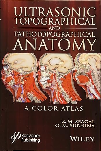
Ultrasonic Topographical And Pathotopographical Anatomy: A Color Atlas PDF is a unique and richly illustrated reference that presents detailed ultrasound anatomy with topographical and pathotopographical perspectives. Designed to guide clinicians in diagnostic ultrasound, this atlas combines high-quality images, anatomical diagrams, and clinical correlations. It is an essential resource for radiologists, sonographers, surgeons, and medical trainees who require precise anatomical visualization for accurate interpretation.
Why This Book Matters
Ultrasound remains one of the most widely used imaging modalities due to its safety, accessibility, and diagnostic value. However, accurate interpretation depends on a clear understanding of anatomical structures in relation to topographical and pathological changes. Ultrasonic Topographical And Pathotopographical Anatomy provides clinicians with comprehensive visual guidance, ensuring precise recognition of both normal anatomy and pathological findings in daily practice.
For trusted updates in imaging, visit the Radiological Society of North America (RSNA) and the European Society of Radiology (ESR).
Key Features of the Ebook
This atlas includes:
-
Full-color ultrasound images with detailed annotations
-
Correlations between topographical and pathotopographical anatomy
-
Practical orientation for diagnostic interpretation
-
Illustrations highlighting normal and pathological findings
-
Coverage of major anatomical regions and organ systems
-
Step-by-step visual guidance for clinical use
-
Updated content reflecting current sonographic standards
For more reference material, consult the American Institute of Ultrasound in Medicine (AIUM) and the journal Ultrasound in Medicine and Biology.
Who Can Benefit
This ebook is designed for:
-
Radiologists and sonographers
-
Surgeons and clinicians using ultrasound in practice
-
Medical students and residents specializing in imaging
-
Physicians in emergency and critical care medicine
-
Educators teaching anatomy and imaging sciences
For complementary resources, explore Atlas of Musculoskeletal Ultrasound Anatomy PDF and Diagnostic Ultrasound 5E PDF.
Learning and Application Strategies
Ultrasonic Topographical And Pathotopographical Anatomy emphasizes visual learning through high-quality ultrasound images and diagrams. Its structured format allows clinicians to quickly locate anatomical regions, compare normal and pathological appearances, and apply findings directly in clinical practice. By correlating topographical anatomy with pathotopographical changes, it supports more accurate diagnoses and improved patient outcomes.
For further educational support, visit the World Federation for Ultrasound in Medicine and Biology (WFUMB) and the British Medical Ultrasound Society (BMUS).
Detailed Content Overview
Chapters are organized to cover:
-
Fundamentals of ultrasound anatomy
-
Topographical anatomy across body regions
-
Pathotopographical anatomy and clinical correlations
-
Organ-based ultrasound imaging
-
Musculoskeletal and vascular anatomy in ultrasound
-
Key pathological examples and diagnostic insights
-
Quick-reference charts and annotated illustrations
Conclusion
Ultrasonic Topographical And Pathotopographical Anatomy: A Color Atlas PDF provides a comprehensive and visually rich approach to understanding ultrasound anatomy. With its unique integration of topographical and pathological perspectives, it serves as a trusted guide for clinicians, educators, and trainees working in diagnostic imaging.
👉 Download Ultrasonic Topographical And Pathotopographical Anatomy: A Color Atlas PDF today to expand your ultrasound expertise. For further reading, visit freemedbooks.com and explore official copies on amazon.com.


