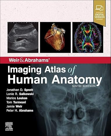
Weir & Abrahams’ Imaging Atlas of Human Anatomy 6E PDF is an authoritative and visually rich reference that integrates advanced imaging techniques with detailed human anatomy. Authored by leading experts, this sixth edition provides high-quality MRI, CT, ultrasound, and X-ray images aligned with precise anatomical illustrations, making it an essential resource for radiologists, anatomists, medical students, and clinicians. With its clear structure and updated imaging content, the atlas enhances both learning and clinical practice.
Why This Book Matters
Accurate anatomical knowledge is fundamental for interpreting diagnostic images and performing clinical procedures. Weir & Abrahams’ Imaging Atlas of Human Anatomy 6E bridges the gap between medical imaging and anatomical study by offering side-by-side comparisons of images and diagrams. This practical approach allows readers to recognize normal anatomy, identify variations, and detect pathology with greater accuracy.
For further authoritative resources, visit the Radiological Society of North America (RSNA) and the British Institute of Radiology (BIR).
Key Features of the Ebook
This imaging atlas includes:
-
Comprehensive coverage of human anatomy using MRI, CT, X-ray, and ultrasound
-
Correlated anatomical diagrams for clear understanding
-
Updated and high-resolution images reflecting current imaging standards
-
Systematic coverage of head, neck, chest, abdomen, pelvis, and limbs
-
Clinical notes highlighting applied anatomy and imaging relevance
-
User-friendly layout with labeled illustrations and concise explanations
-
Expanded content supporting medical education and clinical training
For more academic resources, explore Clinical Anatomy (Wiley) and the Journal of Anatomy.
Who Can Benefit
This ebook is designed for:
-
Medical students preparing for anatomy and radiology exams
-
Radiologists and radiology residents
-
Surgeons and anatomists
-
Clinicians using imaging in daily practice
-
Educators and researchers in medical anatomy and imaging
For complementary titles, see Netter’s Atlas of Human Anatomy PDF and Gray’s Anatomy for Students PDF.
Learning and Application Strategies
Weir & Abrahams’ Imaging Atlas of Human Anatomy 6E emphasizes practical learning by correlating real diagnostic images with anatomical illustrations. This method not only strengthens understanding of anatomy but also sharpens image interpretation skills. Its concise format, systematic chapters, and annotated figures ensure quick reference during academic study, clinical rotations, or radiology practice.
For more insights into imaging and anatomy, visit the European Society of Radiology (ESR) and Clinical Radiology (Elsevier).
Detailed Content Overview
Chapters are organized to cover:
-
Head and neck anatomy with CT and MRI correlations
-
Thoracic imaging including chest radiography and cross-sectional anatomy
-
Abdomen and pelvis with CT, ultrasound, and MRI detail
-
Upper and lower limb anatomy with musculoskeletal imaging
-
Neuroanatomy and cranial imaging correlations
-
Systematic charts and tables for quick review
Conclusion
Weir & Abrahams’ Imaging Atlas of Human Anatomy 6E PDF provides an exceptional integration of anatomy and imaging, making it a valuable reference for students, clinicians, and radiologists. With its detailed content, updated images, and practical approach, it supports both education and clinical application in medical imaging and anatomy.
👉 Download Weir & Abrahams’ Imaging Atlas of Human Anatomy 6E PDF today to advance your anatomical and radiological knowledge. For further reading, visit freemedbooks.com and explore official editions on amazon.com.
Related products